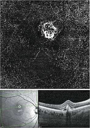
Device aids in diagnosing wet AMD
“Case Scenario,” illustrates how a specific device’s image(s) aided in patient management.
PATIENT
A 60-year-old Caucasian female complained of “a dark spot” OD. Previous OCT B-scan showed a single, small, hard, drusen OD superior-nasal to the fovea.
EXAM FINDINGS
BCVA OD 20/30- OS 20/20. A small, elevated, retinal lesion 1.3 mm superior-nasally to the fovea centralis was seen within a larger area of sub-retinal fluid and coagulated blood. A small, dark scotoma infero-temporally corresponded with the location of the lesion.
APPROACH
Using the Heidelberg Spectralis OCT Angiography (OCTA) software module, a choroidal neovascular membrane in the outer retinal avascular zone was identified. A high resolution B-scan (not shown) showed surrounding sub-retinal fluid and coagulated blood
DIAGNOSIS
Wet AMD; active Type 2 choroidal neovascular membrane
MANAGEMENT
Anti-VEGF injections, which were ultimately successful.
PATIENT CARE TIP
Amsler grid for detection
CODING
H35.3211 wet (exudative) 92014, 92015, 92134
CASE STUDY


SUBMIT YOUR IMAGES AT
bit.ly/CASESCENARIO
Tell us how an image from a specific diagnostic device aided in the care of a patient.


