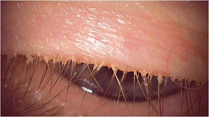
THERE ARE A NUMBER OF DIFFERENT THINGS TO LOOK FOR IN PERFORMING A LID EXAM, SAYS TIM TRINH, O.D., F.A.A.O.
→ ROSACEA. Check the lid margins for redness and inflammation, as this can be a clinical sign of part of a larger facial rosacea. Also, be sure to ask for mask removal to check for facial rosacea.
→ IRREGULAR LID MARGINS. Divots along the lid margins can indicate obstruction and, in some cases, may point to atrophy of the meibomian glands that should be confirmed with meibography.
→ MADAROSIS AND POLIOSIS. Thinning of lashes can indicate chronic blepharitis. Cleaning properly can result in new lashes growing in.
→ PAIN. Press on the lids. Check for pain, and check to see whether the lid margin is stiff. A hardened lid margin can indicate functional blockages.
→ MEIBUM QUALITY. Upon pressing along the lid margins, check for oil consistency. Glands that function well will produce clear oils, whereas those that are obstructed will produce thicker oils. Often, the temporal lid margins can demonstrate more blockages, as these are more prone to be missed when applying compress therapies.
→ KERATINIZATION OF LID MARGINS. Check the surface for a pearly appearance, which indicates keratinization is complete due to chronicity. If the glands are completely blocked, sometimes the only resolution is meibomian gland probing.
→ LID CLOSURE. Have the patient close their eyes behind the slit lamp and assess the lid closures for potential gaps. These gaps can indicate nighttime exposure. OM


