


In their presentation at the 2026 meeting of the American Academy of Optometry, Erika Anderson, OD, FAAO, and Brianne Hobbs, OD, PhD, FAAO, stressed that geographic atrophy (GA) care begins well before a patient develops advanced atrophy or significant central vision loss.
“One of the biggest changes in GA care is that we are learning to recognize the disease trajectory much earlier,” said Dr. Anderson. “Optical coherence tomography (OCT) can show us warning signs years before geographic atrophy becomes clinically obvious or a patient experiences significant central vision loss.”
Dr. Anderson added that optometrists are often the providers who follow these patients over many years. “This gives us the opportunity to recognize structural changes early, document progression longitudinally, educate patients on any newly available therapies, and make timely referrals when appropriate,” she noted.
OCT and FAF
Next, the presenters discussed imaging features on OCT and fundus autofluorescence (FAF) that can help providers identify patients who are at higher risk for progression. The high-risk OCT features they said to look out for are hyper-reflective foci, large drusen volume/height, heterogeneous reflectivity, subretinal drusenoid deposits, and outer retinal disruption.
“For optometrists, the message is that these patients are already in our chairs. Knowing which imaging biomarkers suggest greater risk and following those changes over time gives us an opportunity to be much more proactive in how we counsel and manage them.”
Current and Emerging GA Therapies
Dr. Anderson pointed out that while current treatments have given optometrists the ability to slow geographic atrophy, slowing disease is not the same as restoring vision. “The next phase of GA care will likely involve therapies that target different pathways, reduced treatment burden, and better ways of identifying which patient is most likely to benefit from which approach,” she said.
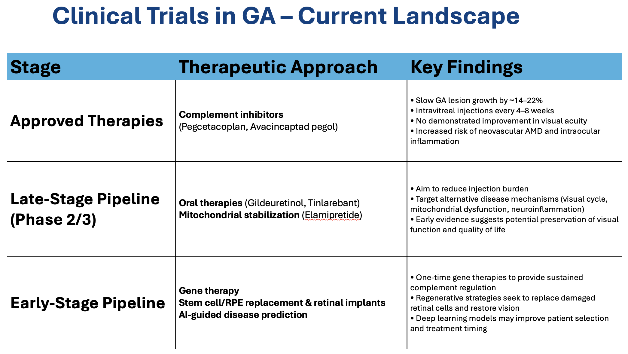
Drs. Anderson and Hobbs then discussed currently available treatments—pegcetacoplan (Syfovre, Biogen) and avacincaptad pegol (Izervay, Astellas)—which are complement inhibitors. They also discussed emerging therapies in the late-stage pipeline—oral therapies and mitochondrial stabilization—as well as early-stage therapies such as gene therapy, stem cell/RPE replacement and retinal implants, and artificial intelligence (AI)-guided disease prediction (Figure 1).
Using AI to Predict Disease Progression
The presenters then discussed the evolving role of AI in predicting disease progression and potentially helping clinicians individualize treatment decisions. They presented an AI-generated slide with real patient progression (from age 59 to 68). The left eye showed real longitudinal imaging from early structural risk to established GA (Figure 2). The analysis showed early high-risk biomarkers that included subretinal drusenoid deposits, outer retinal irregularities, and increasing hyper transmission over time.
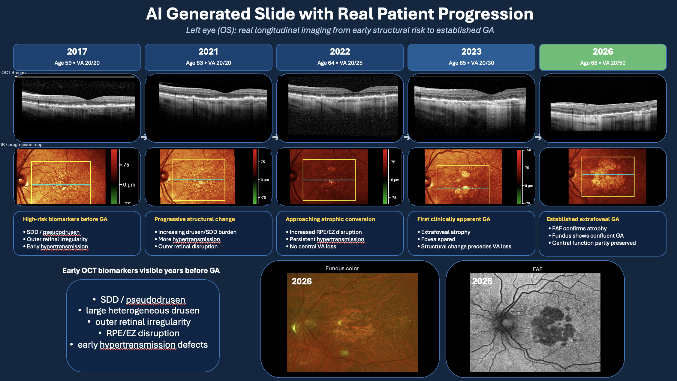
“These were apparent years before GA was clinically apparent,” said Dr. Anderson. “By age 65, extrafoveal GA was present but vision remained preserved. However, at age 68 the GA had progressed into the fovea reducing the visual acuity to 20/50. The case illustrated how structural biomarkers may help identify patients at risk for progression before substantial vision loss occurs.”
Dr. Anderson concluded, “AI is particularly exciting because the goal is not simply to measure an atrophic lesion more accurately. Ultimately, we want to be able to combine longitudinal imaging and clinical information to predict an individual patient's risk of progression, when the fovea may become involved, and eventually what that structural change could mean for their functional vision.” OM