


Visual hallucinations can be a source of significant anxiety for patients who have vision loss, particularly when they fear that what they are seeing indicates a psychiatric disorder, Curt Fritts-Davis, OD, MS, FAAO, said in his “Low Vision, Hallucinations, & Illusions” presentation at the 2026 American Academy of Optometry annual meeting in Anaheim. For optometrists, he said, distinguishing hallucinations from illusions and delusions and understanding other perceptual phenomena associated with low vision can help guide patient education and clinical management.
An illusion is the misperception of a real stimulus
A hallucination is perception in the absence of a stimulus
A delusion is a false belief that cannot be corrected through logic
Charles Bonnet Syndrome
Charles Bonnet Syndrome (CBS) is characterized by visual hallucinations in patients who have vision loss. CBS hallucinations do not occur with other neurologic symptoms, do not involve senses other than vision, are not confused with reality, and do not occur in the absence of vision loss. Their content is also typically not disturbing in nature. What patients see can vary considerably, Dr. Fritts-Davis described, from simple hallucinations—such as photopsias and geometric patterns—to complex images such as animals, people, faces, and other recognizable objects.
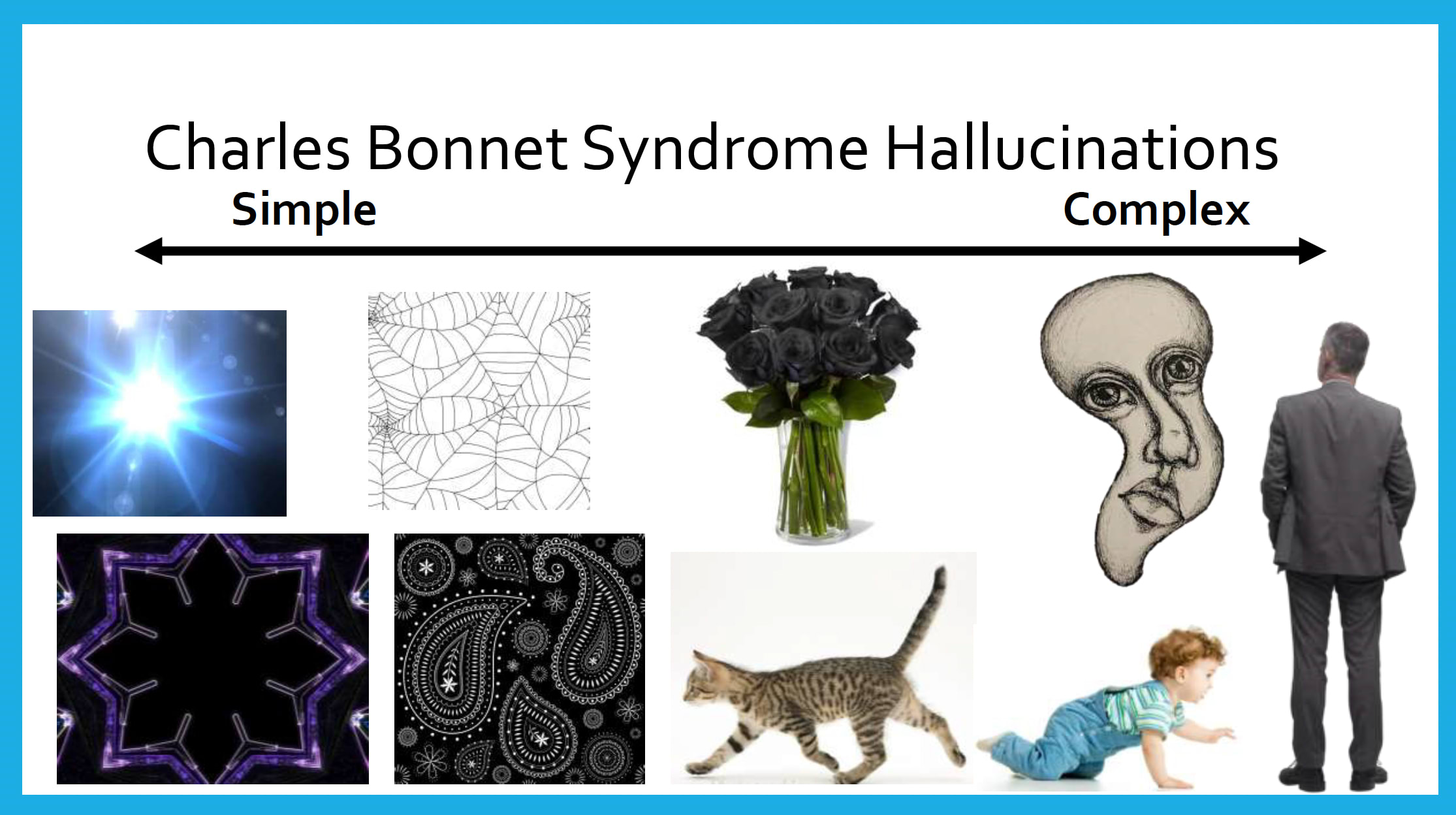
Because patients may be reluctant to volunteer these experiences, Dr. Fritts-Davis recommended considering proactive education rather than waiting for a patient to initiate the discussion. You can introduce the subject indirectly by explaining that other patients who have vision loss sometimes experience unusual visual phenomena, he suggested. For example, say “You don’t have to tell me if this is true for you, but many of my patients…”
For clinical management, he recommended validating the patient's chief complaint, explaining the phenomenon, providing management strategies, and referring for mental health evaluation when appropriate.
Scotomas
Another perceptual phenomenon relevant to low vision is the brain's ability to “fill in” areas of missing vision, Dr. Fritts-Davis described. Scotomas can be filled perceptually with surrounding patterns, colors, and motion, meaning patients may not recognize their own blind spots, even when a scotoma extends to 30°. One degree of visual angle is approximately equivalent to 2 cm at 1 meter, and the physiologic blind spot corresponds to an area approximately 10 × 14 cm at a distance of 1 meter, or about 4 × 5.5 inches at 1 yard.
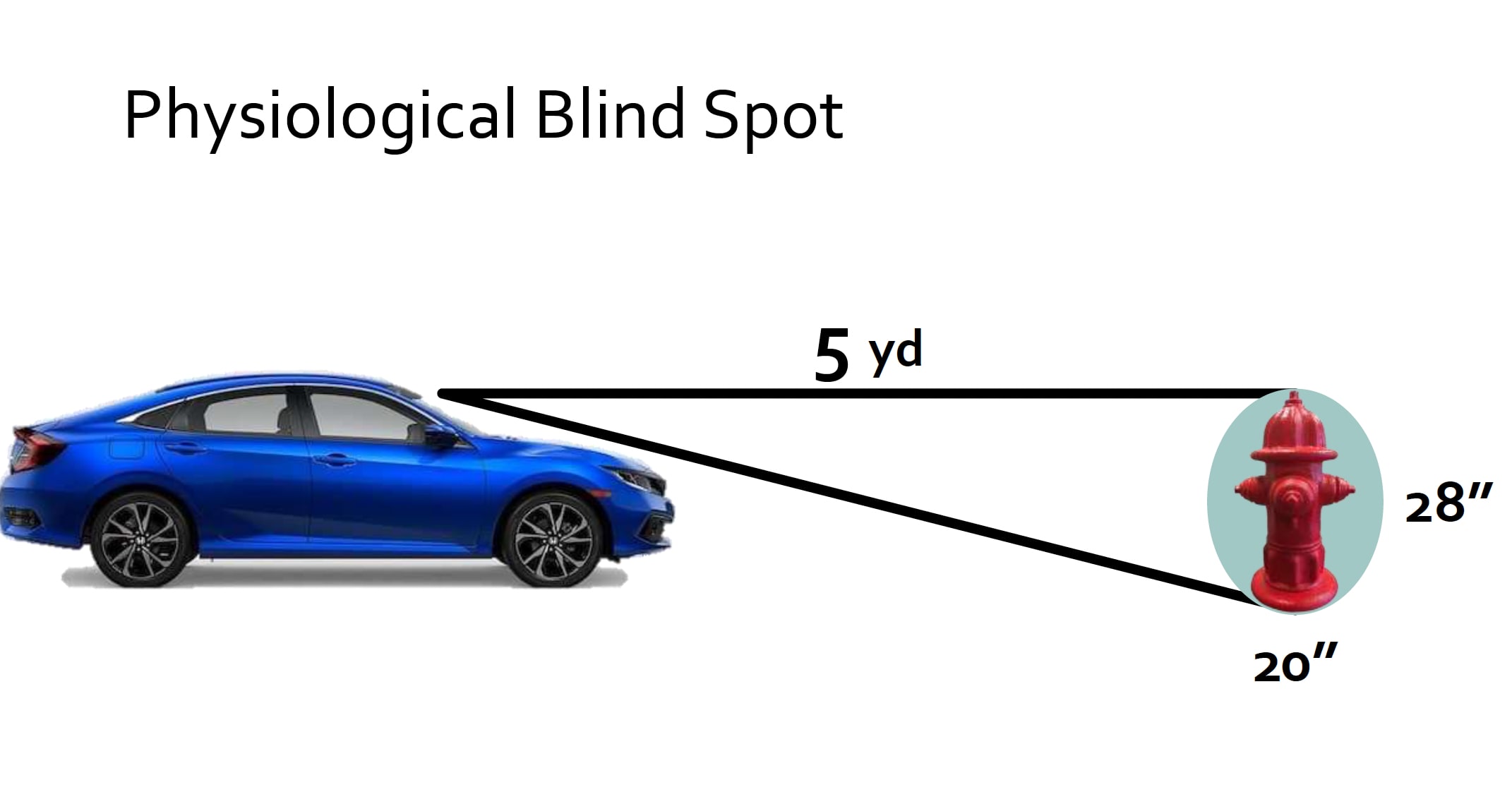
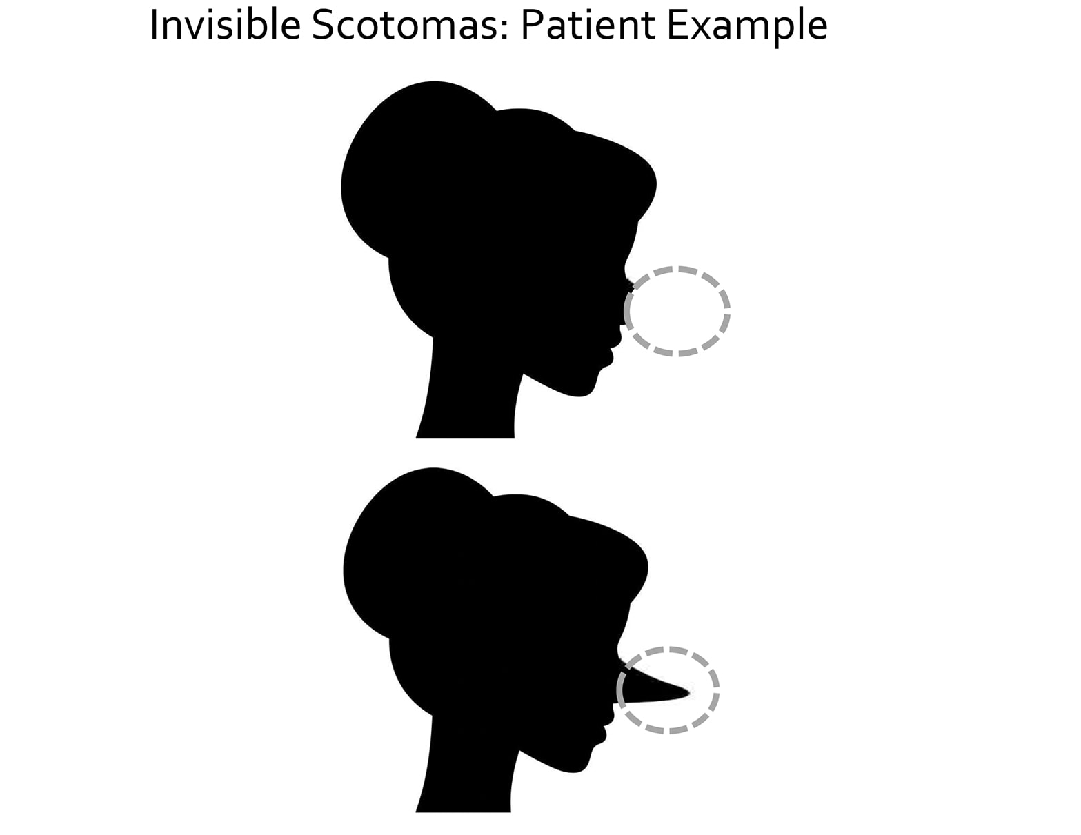
This concept has implications for conversations with patients who have retinal disease and central field loss, he continued. A patient's subjective impression of an intact visual scene does not necessarily mean the visual field itself is intact.
Palinopsia
Low vision patients may also report palinopsia, which is the persistence of vision after a visual stimulus has been removed. Characteristics include an image that retains the same color as the original, is high resolution, and can be long lasting.
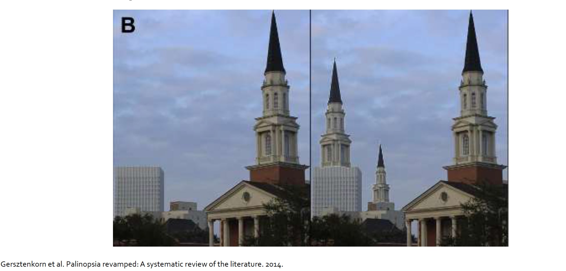
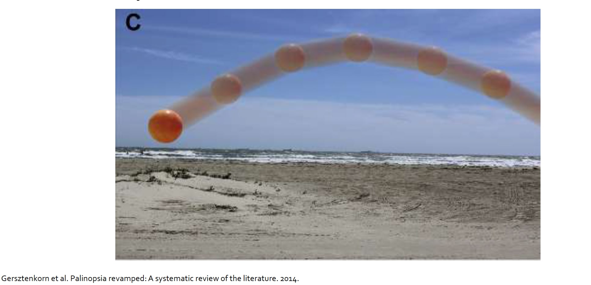
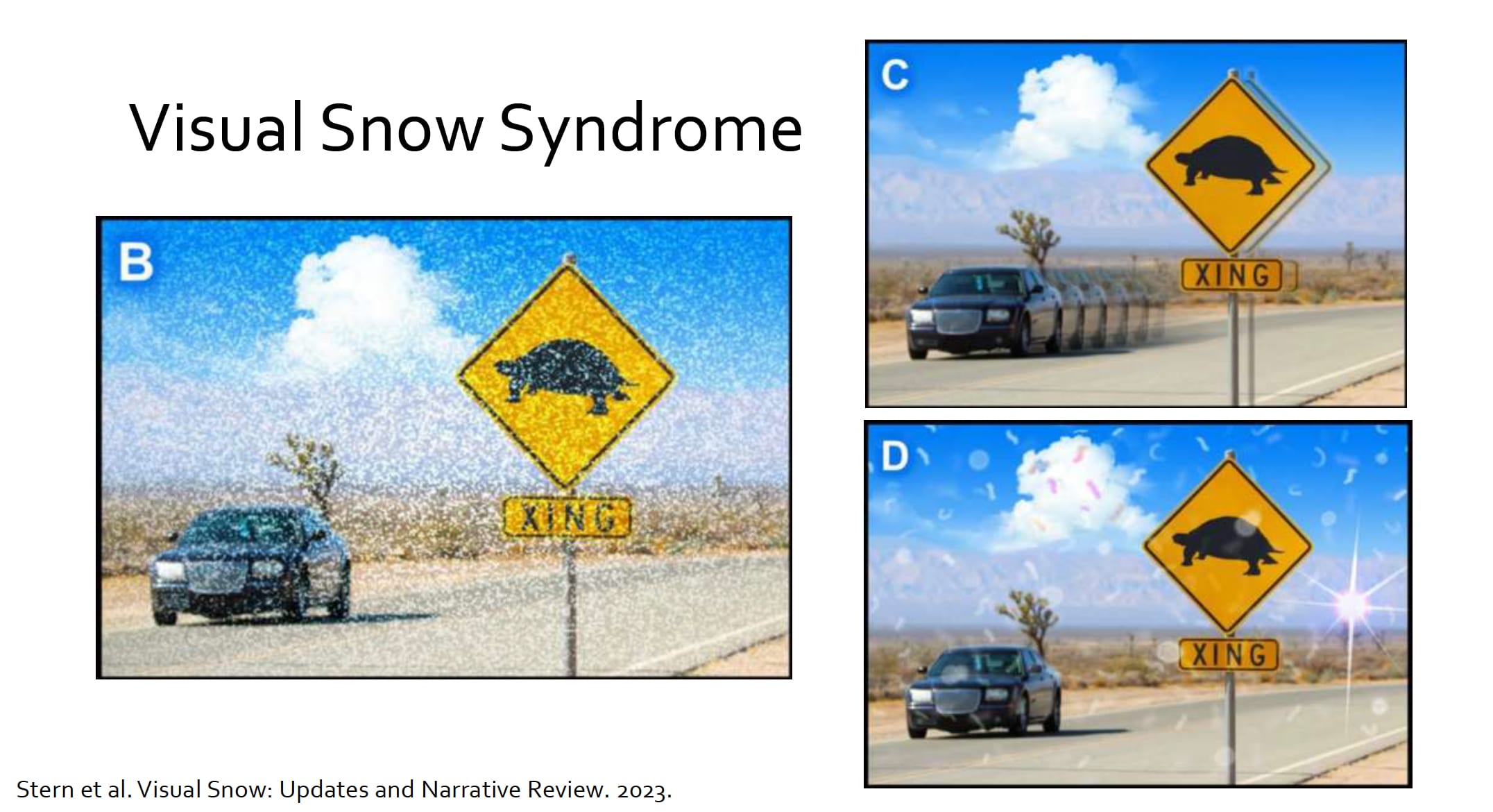
There are several possible etiologies, Dr. Fritts-Davis said, including vision loss associated with deafferentation of the visual cortex; brain damage from trauma, stroke, or a space-occupying lesion; prescription or recreational drug use; and visual snow syndrome. Visual snow syndrome can itself include a group of visual symptoms, such as visual snow, photophobia, palinopsia, nyctalopia, and enhanced entoptic phenomena.
Color Vision Changes
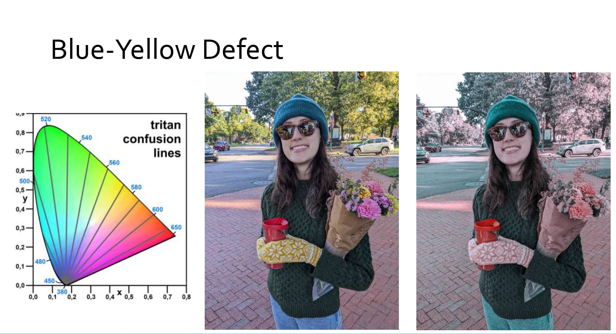
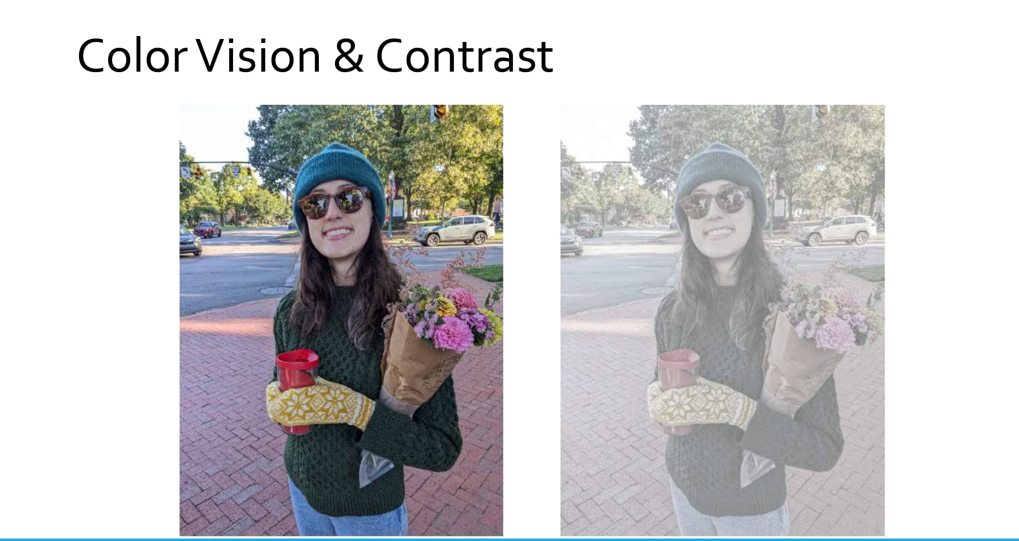
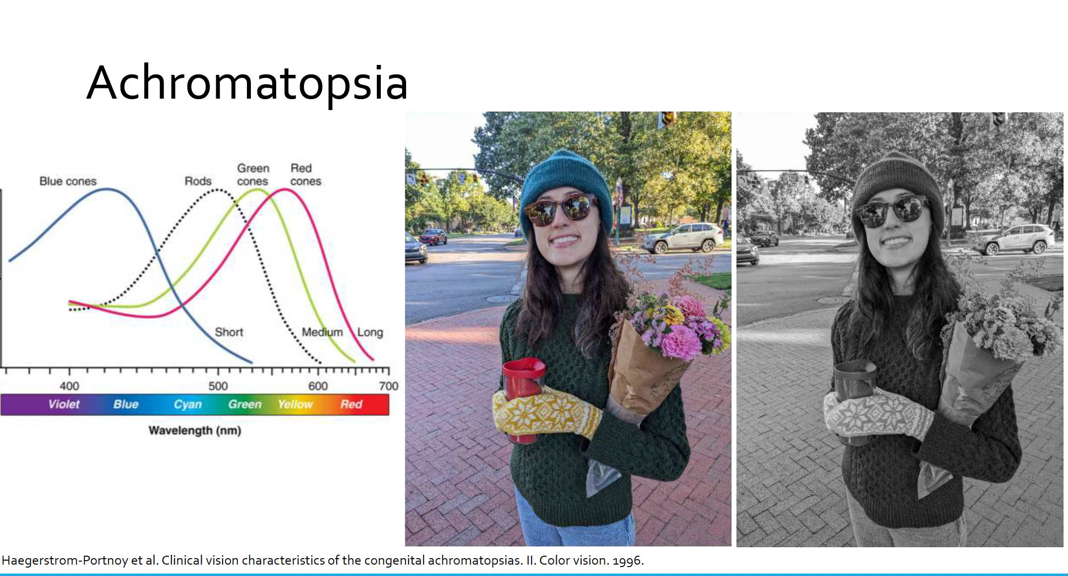
Using the International Commission on Illumination chromaticity diagram, Dr. Fritts-Davis illustrated red-green defects (protan and deutan confusion lines) and blue-yellow defects (tritan confusion lines) to demonstrate how different color defects can alter the appearance of the same visual scene.
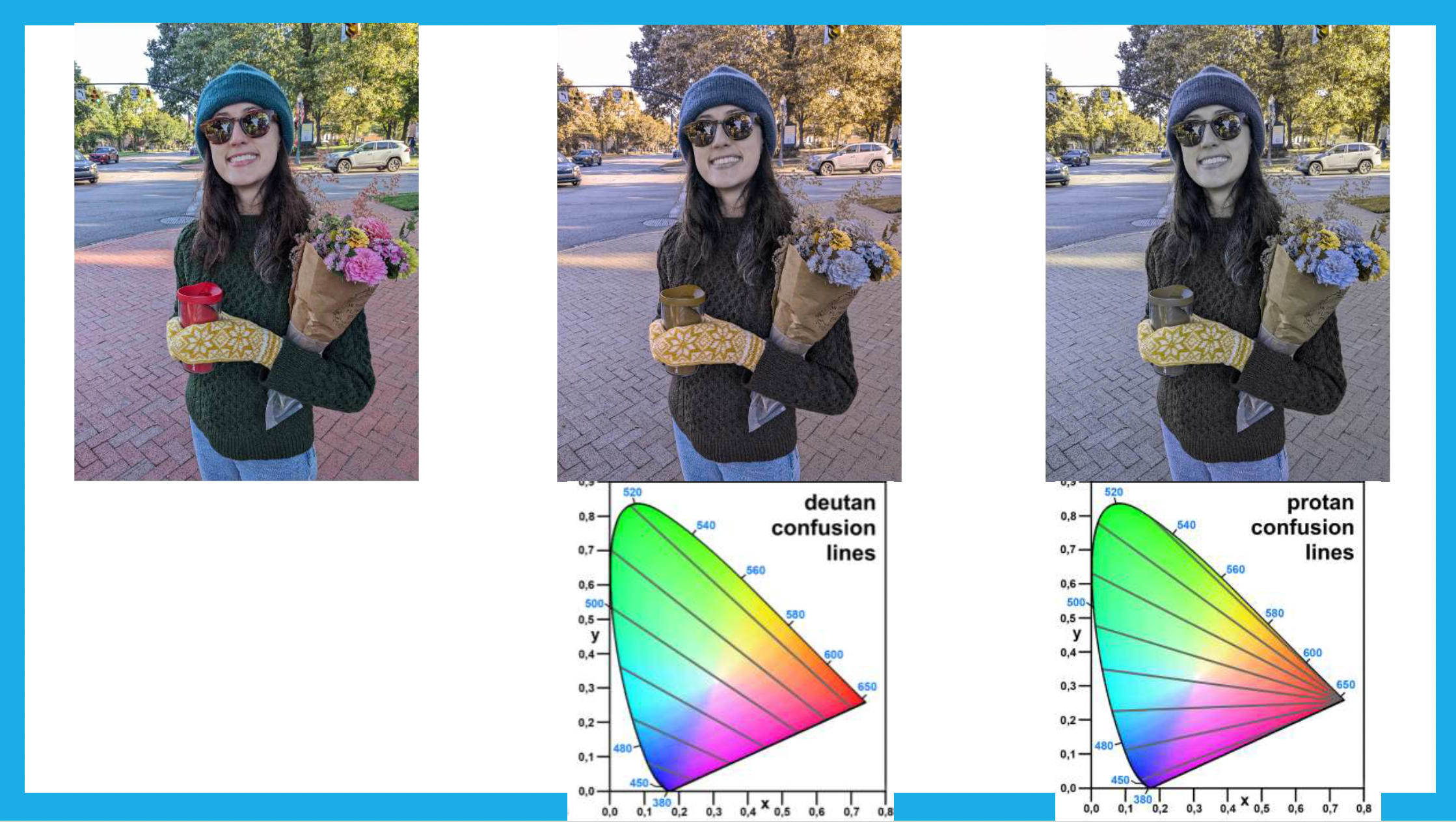
Achromatopsia provided another example of the distinction between color and luminance information. Patients can potentially differentiate stimuli depending on relative brightness or darkness despite markedly impaired color discrimination.
Statokinetic Dissociation and Blindsight
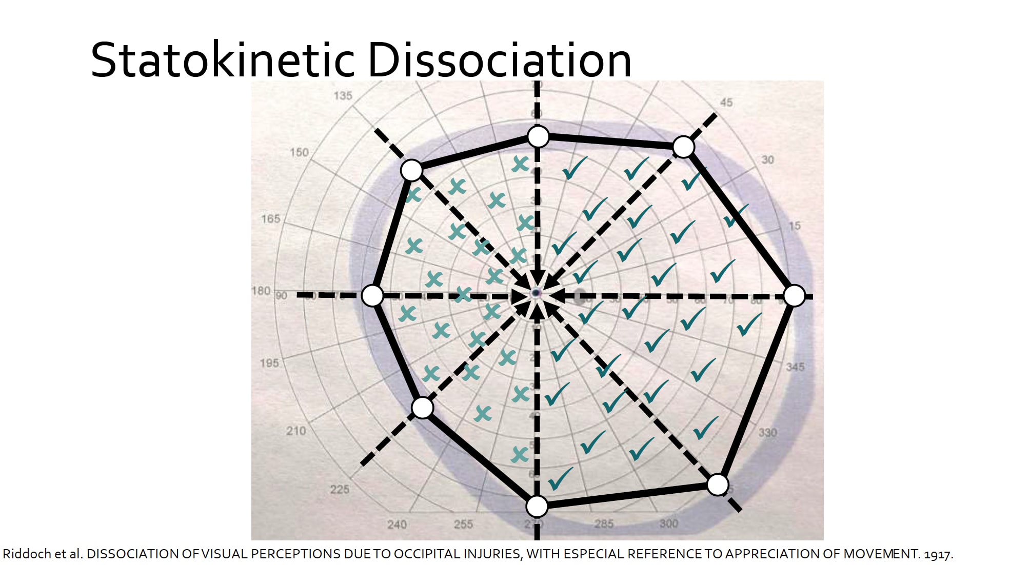
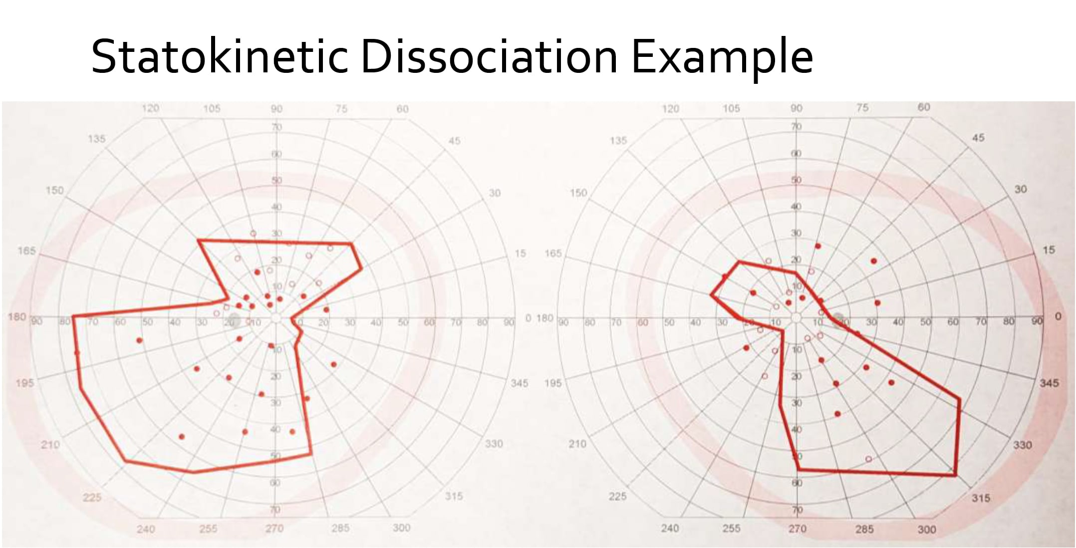
Vision loss can also produce seemingly contradictory findings between measured visual fields and what a patient reports seeing. Statokinetic dissociation, also known as the Riddoch phenomenon, refers to the ability to perceive motion within an otherwise blind visual field. The phenomenon can occur following damage to the primary visual cortex, or V1.
This phenomenon differs from blindsight, Dr. Fritts-Davis said, which is an uncommon phenomenon that is also associated with damage to V1. Patients are perimetrically blind, usually with hemianopia, but can identify shapes, colors, or locations within the blind field at rates better than chance. For example, one blindsight case illustrated the extent of this residual visual processing. Among other testing tasks, the patient correctly identified 89% of animal drawing they were shown in their blind field.
Clinical Takeaways: Ask About Perception in Low Vision Care
Dr. Fritts-Davis emphasized that hallucinations should not be equated with delusions and that both understanding and conveying the distinction may reduce patient anxiety. For optometrists, the phenomena discussed in the session provide additional context for patient reports that may initially appear inconsistent with conventional acuity and visual field findings. "I hope attendees enjoy learning about the distortions to human perception that occur when the visual pathway is damaged, and I hope they can use this information to comfort their patients that may find these abnormal perceptions concerning,” he concluded.