


Optical coherence tomography (OCT) technology has transformed the care of our glaucoma patients dramatically in the past decade. Each innovation in the technology involved in OCT image acquisition has resulted in more information available to us. This is critically important when we are managing complex cases: The more information we have before us, the higher level of patient care we can deliver.
Take, for example, patients who have glaucoma but also have a copresenting optic nerve anomaly unrelated to the glaucoma. The challenge before us essentially becomes twofold: making the diagnosis of glaucoma, and determining how much of what we see structurally is related to glaucoma vs the optic nerve aberration. Is 95% of what we see related to glaucoma (that makes things easy for us), or is, for example, only 50% of what we see with the optic nerve related to glaucoma, and the other 50% is related to the optic nerve anomaly? Issues like high myopia, tilted nerves, and nonglaucomatous optic neuropathies (to name a few) can cloud the picture of glaucoma.
Case Study
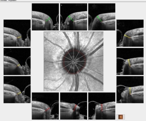
A patient presents with early glaucoma in the both eyes, with no other confounding issues in the right eye, and a significantly vertically tilted optic nerve in the fellow eye. The right eye is easy: There are early structural defects associated with glaucoma, in the form of neuroretinal rim thinning in the inferotemporal quadrant of the optic nerve and confluent retinal nerve fiber layer (RNFL) thinning in the same area. Figure 1 shows the Bruch’s membrane opening-minimum rim width (BMO-MRW) overview of the right eye. Note the relatively plush neuroretinal rim everywhere except for the inferior temporal segment, which typically is where early glaucomatous damage is seen.
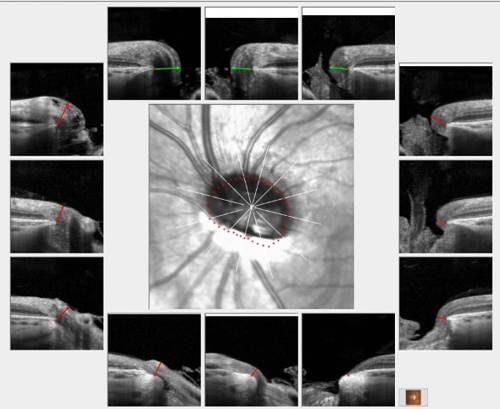
The BMO-MRW overview of the left eye however (Figure 2), shows the significantly tilted insertion of the optic nerve, and aberration of the neuroretinal rim tissue of the inferior half of the optic nerve.
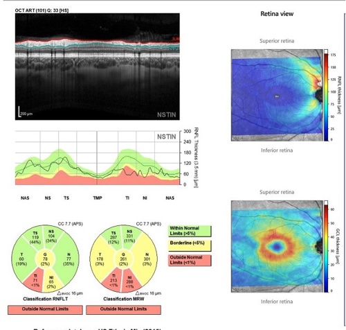
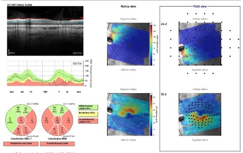
In Figures 3 and 4, we see the Hood reports of the patient’s right and left eyes, respectively, generated from the Spectralis OCT (Heidelberg Engineering). Note in Figure 3 the thinning of the RNFL, which matches the thinning of the neuroretinal rim in the same sector. These findings are consistent with early glaucoma.
In Figure 4, we can see the significant effect to the RNFL thickness measurements owed to the vertically tilted disc. This matches the structural findings seen in the BMO-MRW overview of the same eye. What we do not see here is damage that we can contribute only to glaucoma. Chances are, any glaucomatous damage is hidden by the deformation caused by the tilted disc itself.
We need to keep in mind that, in many situations, patients become their own references insofar as asymmetry is concerned. In this case, there is clear asymmetry, due primarily to the tilted disc. But the right eye clearly shows early damage associated with glaucoma. Given that other metrics observed in the workup of this patient are symmetrical—such as pachymetry, IOP, refractive error, and angle anatomy—it is reasonable to assume that because the right eye has sufficient early damage from glaucoma, the same is probably true of the left eye. Thus, treatment to mitigate further progression of the glaucoma is warranted in both eyes.
Conclusion
Diagnosis of the right eye in this case is straightforward; the left eye not so much. Having detailed information related to both eyes, in the form of advanced OCT imaging, helps us to formulate a specific management plan based on an abundance of structural detail available to us.

This content supported by Heidelberg Engineering, Inc