

Often called “the thief of sight,” glaucoma was estimated to affect 4.22 million people aged 18 years or older in the United States in 2022; of these patients, about 35% had some issue that affected their vision.1 Primary care optometrists check patients daily for glaucoma through intraocular pressure (IOP), optic nerve evaluation, visual field testing, and other components of a comprehensive eye exam. Most of our time is spent on the diagnosis and treatment of glaucoma; consequently, optometrists often focus less attention on its functional complications until later stages. Glaucoma can cause difficulty with reading, driving, visual searching, mobility, and other visually guided activities of daily living,2 which can be present even early in the disease.
Referring Glaucoma Patients for Low-Vision Therapy
Historically, eye diseases like macular degeneration that affect central acuity make up most referrals in low-vision practice.3 Regardless of the type of vision loss, barriers in making a low-vision referral exist for both patients and practitioners. These barriers include patients’ reluctance to referral due to nonacceptance of their vision loss; poor understanding of how low-vision rehabilitation can help; lack of communication with their optometrist about their visual concerns; or health, cognitive, transportation, financial, or other social issues.3,4 Eyecare providers have reported being unfamiliar with referral criteria, available services, and not having enough time to discuss vision rehabilitation in a busy practice.4,5
When it comes to glaucoma, functional visual problems may take a back seat to its medical management, which may be subtle and easily overlooked. In a recent study, Ernst et al showed how often this may happen. Among 522 patients with severe open-angle glaucoma, 88% met the criteria for low vision, but only 2.7% were referred.6 Patients were more likely to be referred when their visual acuity (VA) was reduced, suggesting if central acuity remained relatively good, the need for rehabilitation could be missed.6 In our clinic, we often see glaucoma patients referred only in the later stages of the disease, when VA and visual fields are already compromised. However, many glaucoma patients experience visual difficulties even with relatively preserved visual acuities and fields, which can be addressed at the primary care optometrist’s office.
Careful consideration of patients’ complaints can lead to earlier interventions that support those who have glaucoma. Often, their complaints will include difficulty seeing at night, difficulty with steps, trouble finding items (eg, in a dark bag), and the need for more light.7 Patients with glaucoma may describe these problems even when standard clinical measurements are normal. In some patients, reduced contrast sensitivity may contribute to these functional difficulties, which helps explain why patients may report visual complaints despite good acuity.8
Contrast Sensitivity, Visual Fields, and Visual Function
Contrast sensitivity is important to vision in terms of how well people function in their daily lives, and it can reveal visual problems that may not be apparent on a standard acuity chart. A patient can have 20/20 VA and still have reduced contrast sensitivity.9 This is particularly important in glaucoma, where contrast sensitivity has been shown to predict how well patients perform visually related daily activities. In one study, contrast sensitivity was more strongly associated with performance on these activities than visual field measurements.10
Reading is one of the most common complaints that patients report during a low-vision examination.11 In glaucoma, reading difficulty may be related not only to acuity, but also to visual field loss and reduced contrast sensitivity. These factors have been associated with slower reading and reading fatigue.12,13
There are several methods for measuring contrast: Arden grating, Cambridge low-contrast grating, Pelli-Robson, vision contrast test system (VCTS) Vistech chart, Reagan chart, and FACT chart.14 We use the Pelli-Robson contrast sensitivity chart in our clinic because it uses large letters of consistent size while the contrast between the letters and the background progressively decreases. Higher scores indicate better contrast sensitivity; lower scores indicate poorer contrast sensitivity. However, even without having a specialized chart for testing, easy solutions such as better lighting for reading (task lighting; Figure 1) or yellow color filters (ie, fit-overs; Figure 2) for use in low light can be subjectively tested in the office. A digital chart might already have a way to reduce optotype contrast for subjective testing.
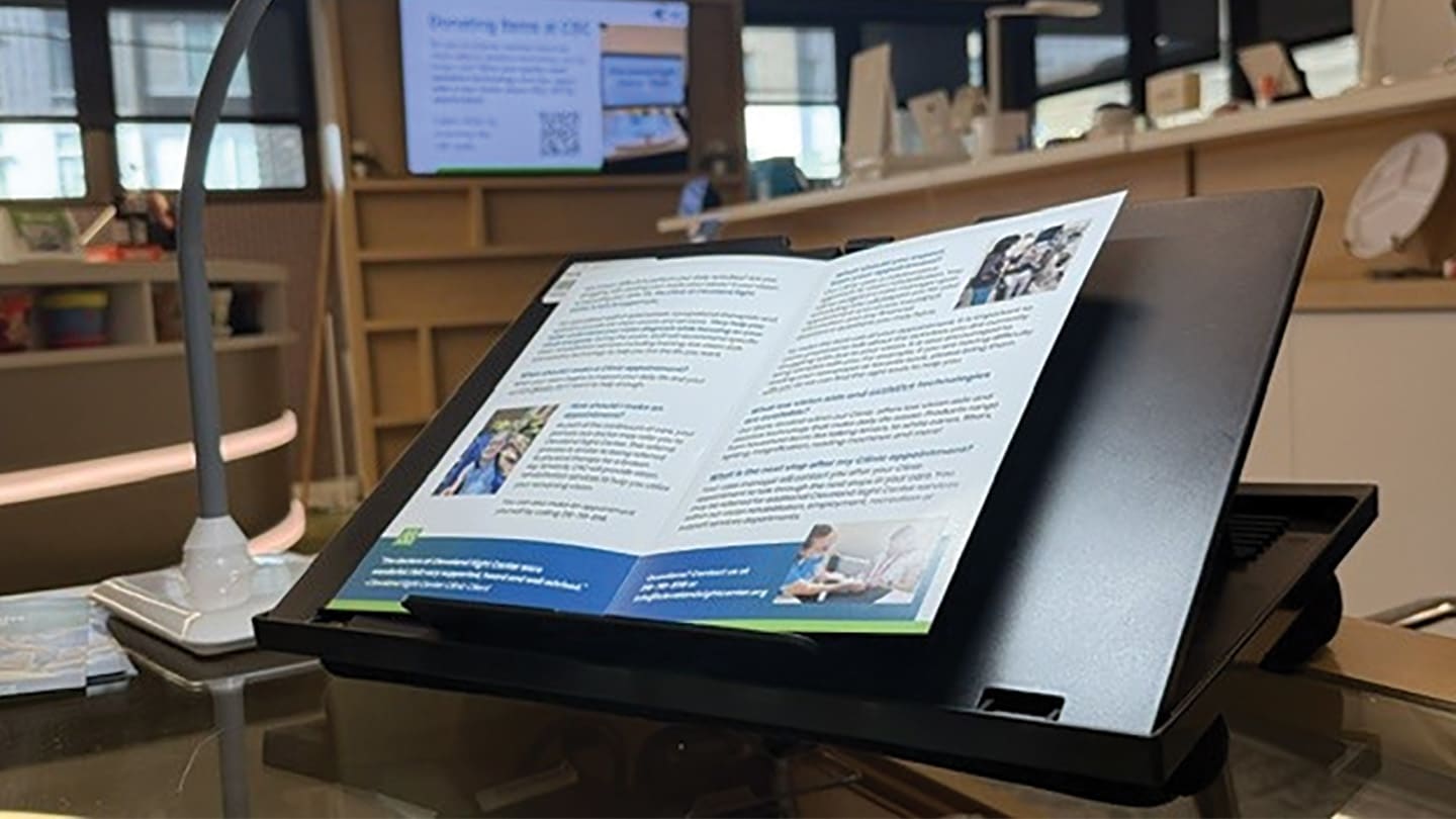
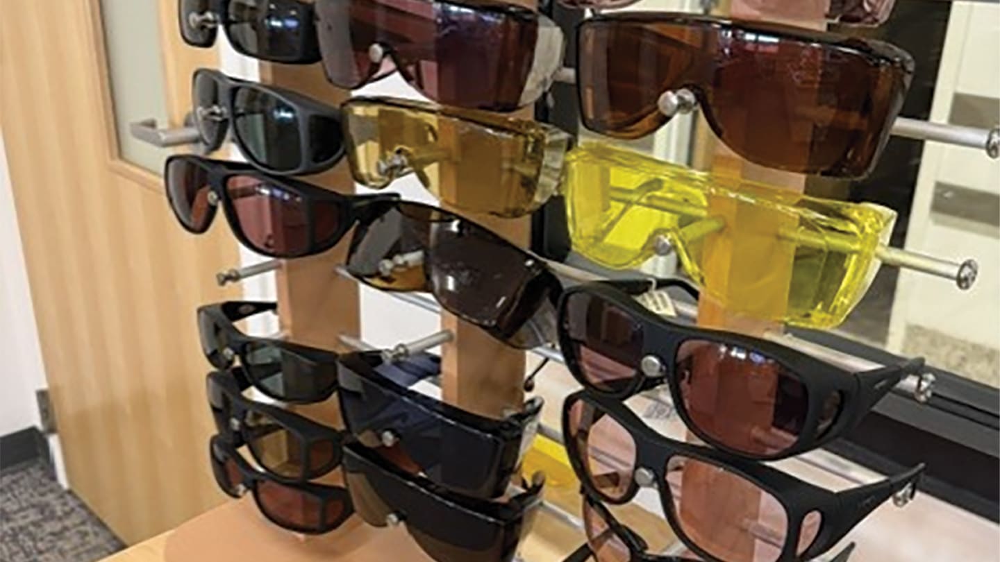
A variety of relatively simple strategies, including improved lighting, appropriate near correction or bifocal design, magnification, and filters or other methods to improve contrast and reduce glare, may help your glaucoma patients both early on and in later stages of the disease. The following cases represent patients seen in our low-vision clinic and highlight practical clinical pearls that may be addressed in the primary care optometrist’s office, as well as in situations in which a low-vision rehabilitation referral or other specialists should be considered.
Case 1: Good Acuity Does Not Mean Good Function
A 69-year-old man with primary open-angle glaucoma (POAG) was referred by his treating eye doctor for a low-vision evaluation. He was using latanoprost OU, and his IOPs were 9 mmHg OD and 9 mmHg OS. His primary complaint was not being able to see small print through his bifocals. Upon further probing he stated that this has been a problem for the past 2 years. He likes to do most of his reading in the den, but now tends to read in the kitchen where the light is better. Additionally, he reported difficulty reading emails on his smartphone and sometimes being sensitive to glare when outdoors during the day.
Entering acuities were 20/25 OD and 20/25 OS with near reading ability of approximately 10-point print OU. His visual fields were also relatively intact. Contrast sensitivity testing was normal.
Trial frame refraction without changes showed that a single-vision near prescription improved subjective reading of small print. Additional lighting with a task lamp (which produces minimal heat during extended use) also subjectively improved reading and helped to increase reading speed. A few simple adjustments to increase font size, brightness, and turning on bold text on his smartphone helped to make reading emails easier. For outdoor glare, he benefited from gray polarized fit-over filters.
For this patient, the solution was not simply additional magnification. His difficulty was related more to lens design and lighting needs. He did well with a single-vision near prescription and the addition of task lighting, which can be positioned where he needs it.
Today, visual function on our smartphones, tablets, and other digital devices should be considered part of our patients’ activities of daily living. Sometimes simple changes to the display settings can make all the difference. Patients in our clinic can be referred to occupational therapy or an assistive technology instructor when more extensive digital needs are required.
Clinical Pearls
- Before increasing the add or prescribing additional magnification, determine what the patient is having difficulty doing. The appropriate treatment may be as simple as providing a dedicated pair of reading glasses with some additional lighting.
- When it comes to digital devices, simple changes in settings can be all that is needed and can even be adjusted by your staff.
- In cases of those with a more significantly affected visual field, increasing the add or magnification reduces the field of vision and working distance, which can make reading more difficult. In these cases, consider an electronic magnification device and try negative contrast.
- When considering lens designs for those with advanced glaucoma, do not automatically also think they require a single-vision lens design. If central vision is not affected, they may do just as well with a progressive. It really depends on the patient’s needs and lifestyle.
Case 2: Look Beyond the Chief Complaint
A self-referred 83-year-old man with POAG presented with difficulty reading his scratch-off lottery tickets. His daughter also wanted to know why he continuously removed his glasses whenever he tried to see up close. History included use of latanoprost, dorzolamide/timolol, and brimonidine. IOPs were 9 mmHg OD and 5 mmHg OS, with a cup-to-disc ratio of 0.80 OU. Visual fields showed a less than 10° visual field OD and a less than 5° visual field OS.
Entering VAs were OD 20/50 and OS 20/50 with ability to read 16-point print at 35 cm. Although the patient removing his glasses to see up close was an unusual behavior to his daughter, findings of a low amount of myopia were consistent with this behavior. Therefore, rather than increasing his near add, we considered the task.
For spot-reading low-contrast materials, such as lottery tickets, a 4x illuminated handheld magnifier provided the additional magnification and lighting he needed. Further history revealed he needed some assistance with accessibility features on his iPad. However, the most important finding had little to do with his lottery tickets or tablet: He was still driving!
Low-vision evaluations give us an opportunity to ask about activities that may not be part of the patient’s original complaint, and driving should be one of them because visual field loss raises concerns about driving safety. This is particularly important in glaucoma where visual field loss has been associated with increased risk of accidents.15 Due to his much-reduced fields and not meeting the state requirements for driving in Ohio, we provided the patient and his daughter with information to sign up for Para-Transit, a ride service for people with disabilities and those who cannot drive due to a medical condition.
These conversations can be difficult for both the patient and family. Be ready to discuss driving as well as mobility concerns and know about alternative transportation resources in your area that may be appropriate, as well as your state’s vision requirements for driving.
Clinical Pearls
The complaint that that brings a patient into your office may not be the only issue. It is the perfect time to probe for more information, not only for driving but also for:
- Compliance with medications,
- Compliance with follow-up visits with their treating eye doctor,
- Other treatments, such as those for dry eye,
- Modern daily living skills, such as use of digital devices, and
- Mobility.
Know your state’s vision driving restrictions and have transportation resources at the ready. This may include ride services for people with disabilities that are run through your own city or county.
Case 3: It’s About Visual Function, Not Acuity on the Chart
A 76-year-old man with advanced glaucoma was referred by his ophthalmologist for a low-vision evaluation. He was taking dorzolamide/timolol OU and latanoprost OU, with IOP of 16 mmHg OD and OS. Surgical history included a trabeculectomy OS in 2014 and an aqueous shunt in 2022. Cup-to-disc ratios were 0.95 OU. Chart review indicated that he had been lost to follow-up for approximately 1 year. Entering acuities were light perception OD and a slow 20/20 OS.
As we walked from the waiting area to the exam room, we noticed he walked slowly and with caution. As we got into the exam room, we asked, “How are you doing? Are you having any trouble walking in unfamiliar places?” Before he could answer, his wife responded, “Oh, he’s fine.” After that, the patient offered few visual complaints.
Based on the VA and few visual complaints, it might appear that he is functioning well and has adapted to being monocular. However, there was more to the story. Further probing found he was having trouble following along in his prayer book at church, particularly in dim lighting. He also admitted to being slow to react to things from his right side and did worry about falling when in unfamiliar places.
Contrast sensitivity testing on the Pelli-Robson chart measured 1.20 logCS, demonstrating reduced sensitivity. Refraction resulted in a new bifocal prescription with a +2.75 add, and with appropriate task lighting, he was able to read 12-point print. We determined that he could use a low-powered 2.5x handheld magnifier with a light while at church for following along in the prayer book. We also instructed him to get closer to the magnifier if he felt he needed more field of vision (Figure 3).
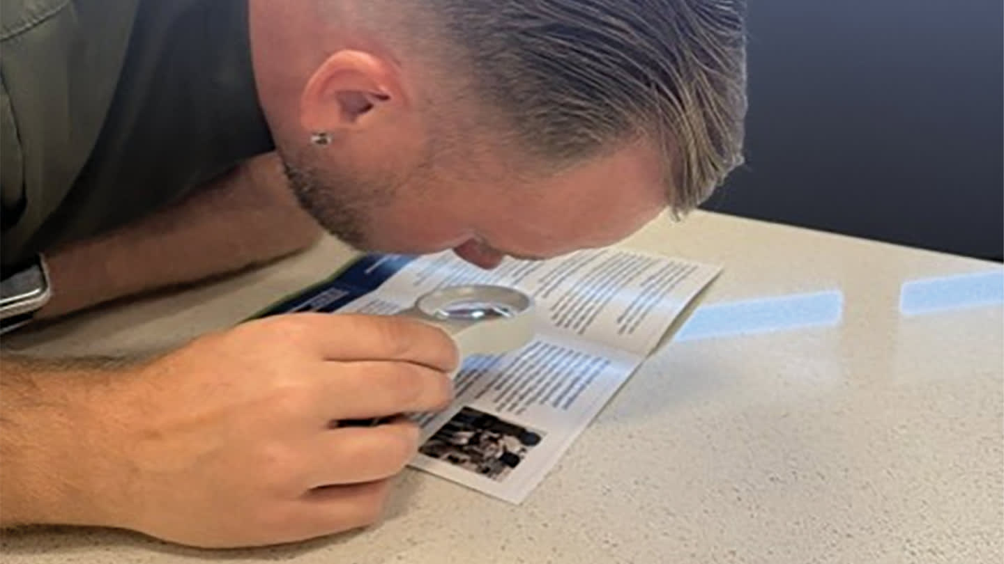
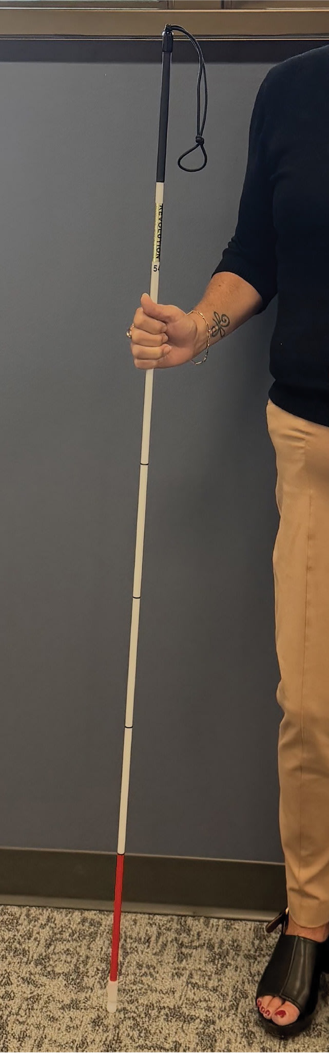
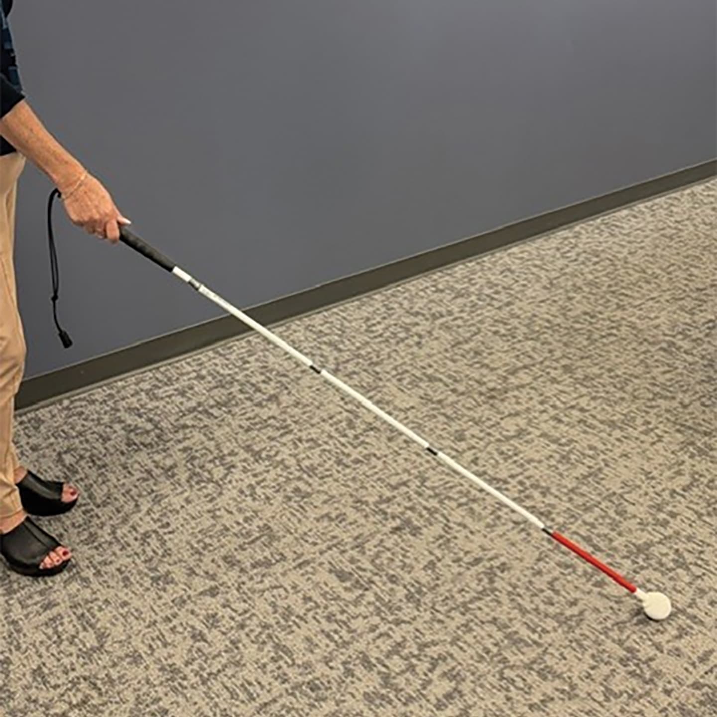
We then discussed scanning techniques for his right side and to consider referral to occupational therapy (OT) for a more complete occupational assessment to make certain other activities of daily living were not impacted. We also discussed possible use of an identification (ID) cane or long white cane for orientation and mobility (Figures 4 and 5).
Clinical Pearls
- Patients and their family members might not understand what a low-vision examination may help with. It is more than just seeing; it is about functioning. Get a thorough history directly from the patient and learn their interests. Although family members want to be helpful, they may only give you part of the story.
- Ask what specific activity the patient is having trouble with, not just whether they are having trouble “seeing.” The right questions can reveal functional difficulties that VA alone may miss.
- Remind patients that getting closer to a magnifier will increase their field of view.
- The knowledge of a patient being lost to follow-up gives the optometrist the opportunity to educate and reinforce, to the patient and their family, the importance of maintaining appointments, as well as adherence to their drops as prescribed.
- OT referral can further reveal any daily living difficulties due to vision loss through a comprehensive occupational profile. This may include problems with shopping, traveling in the neighborhood, taking medications, self-grooming, home cleaning, cooking, and other home-management activities. Although our clinic benefits from being a multidisciplinary practice with in-house OT, you may want to consider partnering with an outside OT as a referral source.
- Bringing up the use of an ID cane or long white cane can be a difficult conversation, but it is important when there are concerns about mobility. A long cane can help detect obstacles, steps, and changes in the walking surface and may help reduce the risk of falls and injury. For patients who are not ready for a mobility cane, an ID cane may also be considered. Although it is not used for obstacle detection, an ID cane can be carried as needed to let others know that the person has a visual impairment. This may be particularly helpful in crowded or unfamiliar environments, where it can signal to others that the individual may need additional space or assistance.
Case 4: Mobility Needs
A 74-year-old woman was referred to our low-vision clinic with advancing glaucoma by her glaucoma specialist. She was using latanoprost and dorzolamide/timolol OU, and brimonidine BID OU. Her IOPs were 12 mmHg OD and 10 mmHg OS. Review of her visual fields showed marked restriction that was greater in the left eye than in the right eye.
Her 2 major complaints were that she disliked switching between distance and near glasses and that it was becoming more difficult and frightening to walk outside at night. She explained that although she receives transportation to and from dialysis, she was concerned about arriving home after dark and navigating from the vehicle to her home, particularly as the days become shorter. She also described her vision as “smoky” and “blurry.”
Entering visual acuities were 20/40 OD and 20/30 OS with the ability to read 12-point print up close. Contrast sensitivity was 1.05 logCS on the Pelli-Robson chart, representing reduced contrast sensitivity, and copies of her visual fields were extremely constricted.
During the assessment, she found a 2.5x illuminated handheld magnifier helpful for reading smaller print. However, magnification was not the primary concern.
We spent considerable time discussing lighting and mobility. A yellow fit-over filter also seemed to subjectively increase contrast of edges of furniture and doorways in the dim-lighted exam room. We talked about making sure that she had adequate illumination when navigating her outdoor stairway, such as using the flashlight on her phone or another light source, and discussed the potential role of a mobility aid. She was not ready to use a long white cane or ID cane, but as in the last case, this conversation opened the door to future discussions. The goal of this case was not simply to improve the smallest print she could read; it was to help her move safely through her environment.
Clinical Pearls
- As in the previous case, bringing up the use of a white cane can be a difficult conversation, but even mentioning the options can open the door to future referrals for orientation and mobility.
- For those with mobility problems at night, consider a small LED flashlight.
- Some patients report subjective benefits from selective tints, such as yellow, at night. Any filter used at night should be trialed under realistic low-light conditions because reducing transmitted light can be counterproductive. Demonstrate in your office with the lights low.
Other Considerations For Filters and Glare
Filters can be helpful for those with glaucoma for indoor, nighttime, and outdoor glare. Often, patients struggle with overhead lighting in stores or in their own home, and ambient lighting can interfere with their ability to navigate. Too dark of a tint can hinder already compromised vision and contrast sensitivity. A light plum tint can be trialed for patients who report discomfort or difficulty with certain indoor lighting, whereas yellow can be helpful for use outside at night.
Outdoor glare can be controlled with amber or gray filters. A polarized filter can reduce glare better than a dark tint. The goal is to reduce glare while preserving as much useful light as possible.
Remember that glare from smartphones and digital devices can also be bothersome. We are optometrists, not tech experts, but the fact remains that smartphone use is now a modern daily living necessity. For glaucoma patients, setting phones to negative contrast (white letters against a black background) may provide more visual comfort. You may also adjust the brightness settings. Sometimes these simple setting changes can make all the difference.
Other Clinical Pearls for Working With Glaucoma Patients and Their Visual Function
- Add: An increased add helps with magnification. However, when a patient has glaucoma, they often need more field of view than magnification. Demonstrate the add with trial lenses in the office before prescribing them, particularly when the patient has significant field loss.
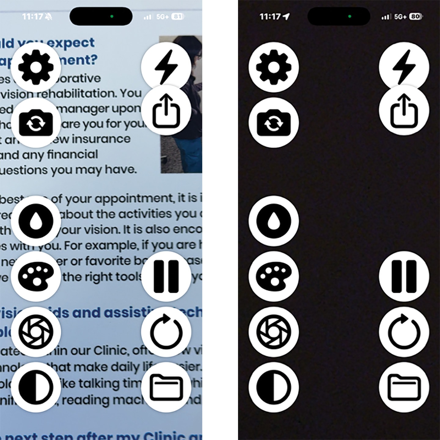
- Handheld magnifiers: Have the patient get closer to the magnifier while maintaining working distance to get the maximum field of view. Also consider electronic magnification with negative contrast or a smartphone magnification app like Rebokeh (available both on iOS and Android; Figure 6) which can adjust color and contrast.
- Photochromics: Many patients do not want to switch between glasses, so photochromic lenses can be convenient for outdoor use. However, some glaucoma patients may have difficulty adapting between bright and dim environments and therefore may find photochromic lenses uncomfortable or disorienting. In these situations, a fit-over filter that can be removed quickly may give the patient greater control over changing lighting conditions.
- Lighting: Make sure lighting does not create veiling glare. It should target the patient’s specific task. Daylight lamps create light that is easy for the eye to use. A lamp that provides adequate light directly to the task may be more useful than increasing overall room brightness. Many also do not get as hot as an incandescent bulb for better safety.
Other Helpful Tips
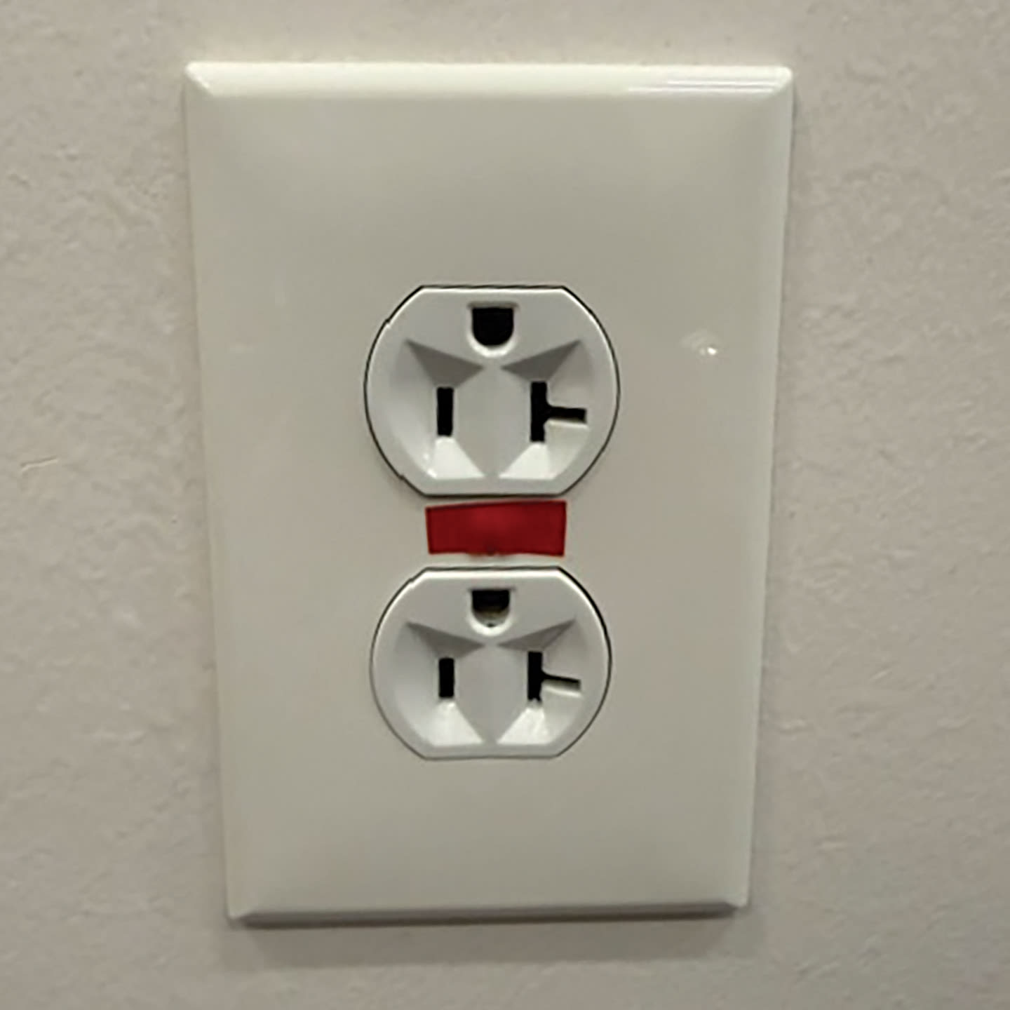
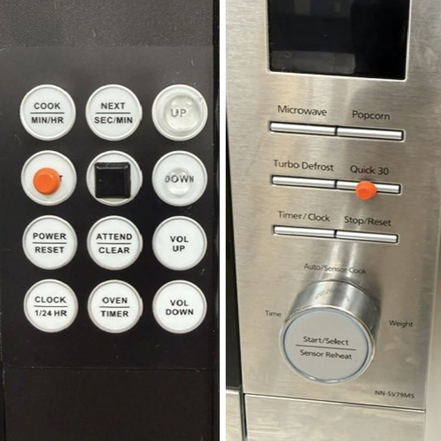
- Increase contrast with labeling: Yellow or red electrical tape, nail polish, or color bump dots can help people find outlets, switches, or buttons on devices such as remotes (Figures 7 and 8). The goal is not to make the object brighter, but to create a stronger color contrast between the object and its background. Bump dots also give a tactile solution for flat panel displays, like on a microwave, or on a television remote to easily find a button.
- Progressive lenses: In low vision, we often prescribe single vision or bifocals to make the most out of the lens. However, we have found that progressive lenses may be preferred by patients who have fairly good central vision, mostly in cases where they wore them previously.
- Dry eye: Consider and treat dry eye secondary to glaucoma medications as a cause of functional visual complaints.
- Coding: ICD 10 has the code H53.72 Impaired contrast sensitivity, which can be used in conjunction with the H40 glaucoma codes. H54 codes (low-vision coding) can be used for visual fields of less than 20°.
Conclusion
Eyecare providers treating glaucoma should routinely ask their patients about their functional vision, even with relatively good acuities and little or no visual field loss. Simple questions may uncover difficulties with reading, lighting, glare, mobility, driving, or even use of digital devices. Some of these problems can be addressed in the primary care office or may benefit from referral to a low-vision provider. Improved lighting, filters, appropriate lens design, accessibility features in our tech, and attention to mobility and safety can make a difference in our patients’ daily lives. Early recognition of functional difficulties in glaucoma allows us to provide patients with early resources and strategies that become more important if the disease progresses. OM
References
- Ehrlich JR, Burke-Conte Z, Wittenborn JS, et al. Prevalence of glaucoma among US adults in 2022. JAMA Ophthalmol. 2024;142(11):1046-1053. doi:10.1001/jamaophthalmol.2024.3884
- Deemer AD, Goldstein JE, Ramulu PY. Approaching rehabilitation in patients with advanced glaucoma. Eye (Lond). 2023;37(10):1993-2006. doi:10.1038/s41433-022-02303-z
- Owsley C, McGwin G Jr, Lee PP, Wasserman N, Searcey K. Characteristics of low-vision rehabilitation services in the United States. Arch Ophthalmol. 2009;127(5):681-689. doi:10.1001/archophthalmol.2009.55
- Stolwijk ML, van Nispen RMA, van der Ham AJ, Veenman E, van Rens GHMB. Barriers and facilitators in the referral pathways to low vision services from the perspective of patients and professionals: a qualitative study. BMC Health Serv Res. 2023;23(1):64. doi:10.1186/s12913-022-09003-0
- Khimani KS, Battle CR, Malaya L, et al. Barriers to low-vision rehabilitation services for visually impaired patients in a multidisciplinary ophthalmology outpatient practice. J Ophthalmol. 2021;2021:6122246. doi:10.1155/2021/6122246
- Ernst J, Huang J, Tsosie J, Ramsey DJ. Factors associated with referral to low vision for patients with advanced glaucoma. Life (Basel). 2025;16(1):12. doi:10.3390/life16010012
- Hu CX, Zangalli C, Hsieh M, et al. What do patients with glaucoma see? visual symptoms reported by patients with glaucoma. Am J Med Sci. 2014;348(5):403-409. doi:10.1097/MAJ.0000000000000319
- Hawkins AS, Szlyk JP, Ardickas Z, Alexander KR, Wilensky JT. Comparison of contrast sensitivity, visual acuity, and Humphrey visual field testing in patients with glaucoma. J Glaucoma. 2003;12(2):134-138.
- Sabel BA, Wang J, Cárdenas-Morales L, Faiq M, Heim C. Mental stress as consequence and cause of vision loss: the dawn of psychosomatic ophthalmology for preventive and personalized medicine. EPMA J. 2018;9(2):133-160. doi:10.1007/s13167-018-0136-8
- Richman J, Lorenzana LL, Lankaranian D, et al. Importance of visual acuity and contrast sensitivity in patients with glaucoma. Arch Ophthalmol. 2010;128(12):1576-1582. doi:10.1001/archophthalmol.2010.275
- Brown JC, Goldstein JE, Chan TL, Massof R, Ramulu P, Low Vision Research Network Study Group. Characterizing functional complaints in patients seeking outpatient low-vision services in the United States. Ophthalmology. 2014;121(8):1655-62.e1. doi:10.1016/j.ophtha.2014.02.030
- Ramulu PY, Swenor BK, Jefferys JL, Friedman DS, Rubin GS. Difficulty with out-loud and silent reading in glaucoma. Invest Ophthalmol Vis Sci. 2013;54(1):666-672. doi:10.1167/iovs.12-10618
- Kwon M, Liu R, Patel BN, Girkin C. Slow reading in glaucoma: is it due to the shrinking visual span in central vision? Invest Ophthalmol Vis Sci. 2017;58(13):5810-5818. doi:10.1167/iovs.17-22560
- Pesudovs K, Hazel CA, Doran RML, Elliott DB. The usefulness of Vistech and FACT contrast sensitivity charts for cataract and refractive surgery outcomes research. Br J Ophthalmol. 2004;88(1):11-16. doi:10.1136/bjo.88.1.11
- Kwon M, Huisingh C, Rhodes LA, McGwin G Jr, Wood JM, Owsley C. Association between glaucoma and at-fault motor vehicle collision involvement among older drivers: a population-based study. Ophthalmology. 2016;123(1):109-116. doi:10.1016/j.ophtha.2015.08.043