


Patients with conditions that lead to poor vision, such as keratoconus, can easily be missed for other important diagnoses if their reduced acuity is automatically blamed on the longstanding pathology. However, a clinician who carefully examines the entire ocular condition can catch a meaningful diagnosis, especially in postsurgical cases.
A 65-year-old man presented 3 months after having cataract surgery in his right eye. His vision had been poor since surgery, and the improvement he expected never occurred. He assumed healing would take time, but after 3 months without meaningful progress, it was clear that this was not an ordinary recovery.
The patient had mild keratoconus and had previously depended on a rigid gas-permeable (RGP) contact lens for his best vision. He had not worn a contact lens in the operated eye since surgery. The initial hypothesis, therefore, seemed straightforward: Cataract extraction had removed the lenticular opacity, but irregular corneal optics remained. A properly designed rigid lens should neutralize much of the anterior corneal irregularity and restore his previous level of vision.
The examination, however, told a different story.
When the Obvious Diagnosis Fails
Before cataract surgery, the patient’s best-corrected visual acuity (BCVA) in the right eye had been 20/40. At the 3-month post-op presentation, it was 20/100. More importantly, a diagnostic RGP fitting failed to produce acceptable acuity, and pinhole testing over the trial lens provided no meaningful improvement.
Usually, a rigid contact lens creates a regular refracting surface over an irregular anterior cornea, with the post-lens tear layer compensating for much of the distortion. Although it cannot eliminate every source of visual degradation in keratoconus—including posterior corneal aberrations, scarring, and residual higher-order aberrations—it should ordinarily provide at least some improvement when anterior corneal irregularity is the principal cause.
The absence of improvement required us to reconsider the diagnosis.
For reduced acuity after cataract surgery, the list of differential diagnoses is long. Poor postoperative vision could potentially arise from residual refractive error, irregular astigmatism, ocular surface disease, corneal edema, posterior capsular opacification, macular disease, optic neuropathy, cystoid macular edema, or an intraocular lens (IOL) that is faulty, tilted, decentered, unstable, or otherwise improperly positioned. In this case, the existing diagnosis of keratoconus created a risk of anchoring bias. The cornea was abnormal, but that did not establish that it was responsible for the magnitude of the new visual deficit.
Slit-lamp examination gave a clue worth investigating. The posterior chamber IOL appeared potentially decentered, although its orientation could not be confidently established from the slit-lamp view alone. Pharmacologic dilation was deferred during the initial evaluation, and we turned to high-resolution anterior segment imaging to determine whether the apparent asymmetry represented a meaningful positional abnormality.
Moving From Impression to Anatomic Evidence
We evaluated the eye using the ANTERION imaging platform (Heidelberg Engineering). It combines swept-source optical coherence tomography (SS-OCT) imaging with anterior segment measurements, allowing evaluation of the cornea, anterior chamber, and implanted lens within a connected imaging environment.
The cornea and cataract apps helped evaluate whether the visual loss could reasonably be explained by corneal geometry or a simple postoperative refractive discrepancy. This distinction is particularly important in keratoconus, where abnormal anterior curvature may appear to be the most important consideration, but posterior corneal shape, total corneal power, axial measurements, and internal optics may have meaningfully changed due to undergoing surgical incisions. In the case of this patient, his keratoconus appeared stable compared to metrics acquired before his cataract surgery referral.
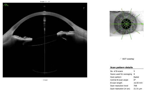
The imaging app on the platform provided more decisive information. High-resolution SS-OCT cross sections enabled direct assessment of the implanted lens. The images confirmed that the IOL was tilted.
This finding unified the clinical picture. The patient’s keratoconus remained stable and visually significant, but it did not explain why his vision was substantially worse than before cataract surgery, or why a diagnostic RGP lens failed to improve acuity. A tilted IOL creates an internal optical abnormality that a corneal contact lens cannot neutralize. Depending on its magnitude and direction, tilt may induce defocus, astigmatism, coma-like higher-order aberrations, glare, monocular diplopia, and loss of BCVA. Pinhole testing may also fail when the retinal image is degraded by significant aberration rather than simple spherical refractive error.
Technology as a Test of the Hypothesis
Technology is most useful when integrated into clinical reasoning, not when substituted for it. The diagnostic process still began with history, acuity, refraction, contact lens optics, pinhole response, and biomicroscopy. The imaging platform became pivotal because it answered a focused question that conventional examination could not resolve confidently: Was the IOL positioned as intended?
The case also demonstrates why a known diagnosis should never become a universal explanation. Keratoconus can account for blurred vision, irregular astigmatism, and dependence on rigid lenses, but it should not automatically explain every postoperative complaint. When the patient’s response does not match the predicted response—particularly when a well-applied diagnostic lens fails to improve vision—that discrepancy should be investigated.
In this case, the ANTERION platform was the investigative tool that allowed for a confident diagnosis. The result was a shorter path to the appropriate specialist, greater confidence in the referral, and a clearer explanation for the patient.

This editorially independent content is sponsored by Heidelberg Engineering