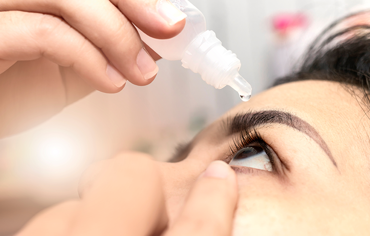
As laser privileges continue to expand across the United States, more optometrists are incorporating in-office laser procedures such as selective laser trabeculoplasty (SLT), laser peripheral iridotomy (LPI), and yttrium-aluminum-garnet (YAG) into patient care. Nicholas R. Green, OD, MPH, FAAO, described the advantages in his “Tips, Tricks, and New Technologies for In-Office Laser Procedures” presentation at Optometry’s Meeting 2026 in Phoenix.
You can find Dr. Green's clinical pearls for SLT here and LPI here.
“With scope of practice laws expanding and an ever-increasing patient need, it is more important now than ever before for optometrists to understand and be comfortable performing laser procedures,” Dr. Green told OM. “Best practices and new technologies are rapidly changing, and it is important for all ODs to be up to date to best care for our patients."
Dr. Green’s Clinical Pearls for YAG Capsulotomy
- Check that the laser is on YAG mode before shooting.
- Be flexible with power and offset settings—offset can be decreased to treat more difficult membranes.
- Try not to have pieces of membrane break free.
- Start treating at 6 o’clock to allow for fluid drainage in posterior capsular distention syndrome.
- Check that the visual axis is clear before finishing the procedure.
YAG Capsulotomy
Posterior capsule opacification remains one of the most common late complications of cataract surgery: Dr. Green noted that approximately half of cataract patients will require YAG capsulotomy at some point. The procedure uses a plasma microexplosion to create shockwaves that dissect opacified posterior capsule tissue and restore visual clarity.
Indications include symptomatic vision loss, glare, refractive shifts in pseudoaccommodating intraocular lenses, obscured retinal views, and preparation for LASIK enhancement. Contraindications include cystoid macular edema, high risk of retinal detachment, calcified intraocular lenses, and ongoing anti-VEGF therapy.
In preoperative planning, the clinician should document pupil size under photopic and mesopic conditions, fully dilate the pupil with tropicamide and phenylephrine, and instill antiglaucoma medication 30 to 60 minutes before treatment. Initial laser settings should be set up to 3.0 mJ, depending on opacification density, single shot, 100 to 250 µm in posterior offset for posterior capsulotomies, and 0 to 100 µm in anterior offset for anterior capsulotomies. The eye should be anesthetized and can be stabilized using a laser lens.
Dr. Green presented a few different techniques, including the continuous ascending linear shot pattern from a recent case series that demonstrated successful improvement in visual acuity with minimal side effects.1
Clinicians can consider prescribing topical steroids BID-QID and antiglaucoma drops if needed, he said. Potential complications include intraocular lens pitting, which reduces image quality, increased floaters, cystoid macular edema, posterior vitreous detachment, vitreous prolapse, corneal burns, and retinal detachment, especially in younger patients. Postoperative follow-up should be at 1 week if the patient has been prescribed steroids, or at 1 month for dilated fundus exam and possible refraction.
Evolving Technology, Expanding Opportunities
Dr. Green concluded that patient selection remains the key determinant of procedural success. As technologies such as direct SLT, micropulse laser trabeculoplasty, and pattern-scanning systems continue to evolve, optometrists are likely to encounter increasing opportunities to integrate laser procedures into clinical practice. Staying informed about current evidence, emerging technologies, and evolving best practices remains essential, he said, whether optometrists perform these procedures themselves or comanage patients who receive them.
Dr. Green has no disclosures to report.
References
-
Lytvynchuk L, Ouali MA, Stieger K, Saksonov S, Bilynska TS, Lumi X. Introducing a modified neodymium:yttrium aluminum garnet posterior capsulotomy technique: case series study of a continuous ascending linear shot pattern. J Int Med Res. 2026;54(3). doi: 10.1177/03000605261416673